Very Short Answer Type Questions
Question. Name the pouch in which the human testes are present.
Answer : Scrotum.
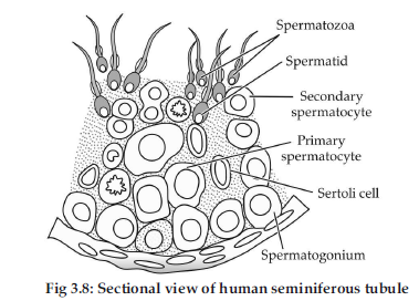
Question. Name the cells that nourish the germ cells in the testes. Where are these located in the testes ?
Answer : Name of cells—Sertoli cells.
Location—They are found lining-up the inner surface of seminiferous tubules.
Question. Write the location and function of the Sertoli cells in humans.
Answer : In the seminiferous tubules of testis.
Function : Nourishes the sperms or germ cells.
Question. Write the location and function of Leydig cells in humans.
Answer : Leydig cells are present at interstitial region between seminiferous tubules. They synthesize and secrete androgens.
Question. Mention the function of trophoblast in human embryo.
Answer : Trophoblast helps to provide nutrition to the embryo.
Question. How does the sperm penetrate through the zona pellucida in human ovum?
Answer : The sperm penetrate through the zona pellucida by releasing sperm lysins.
Question. Name the embryonic stage that gets implanted in the uterine wall of human female.
Answer : Blastocyst.
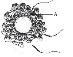
Question. Identify the figure below and the part labelled “A”.
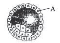
Answer : The given figure is of blastocyst and the part labelled ‘A’ is trophoblast.
Question. How is the entry of only one sperm and not many ensured into an ovum during fertilisation in humans?
Answer : Depolarisation of the egg plasma membrane by binding of sperm to it checks additional sperms from entering into it.
Question. Mention the function of zona pellucida.
Answer : During fertilisation, a sperm comes in contact with the zona pellucida layer of ovum which induces changes in the membrane that block the entry of additional sperms. So, zona pellucida ensures that only one sperm can fertilise an ovum.
Short Answer Type Questions
Question. Name the embryonic stage that gets implanted in human female. Explain the events that occur during this process.
Answer : Blastocyst gets implanted in human female. In a blastocyst, the blastomeres are arranged into an outer layer called trophoblast and an inner group of cells called the inner cell mass. The trophoblast then gets attached to the endometrium and the inner cell mass gets differentiated as the embryo. After attachment the uterine cells divide rapidly and cover the blastocyst. As a result, the blastocyst becomes embedded in the endometrium of the uterus. This whole phenomenon is called implantation and it leads to pregnancy.
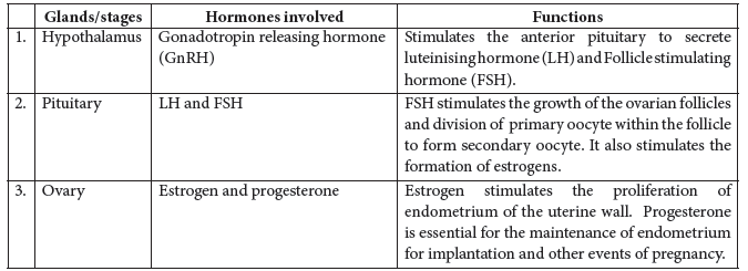
Question. Study the flow chart given below. Name the hormones involved at each stage and explain their functions.
Hypothalamus
↓
Pituitary
↓
Ovary
↓
Pregnancy
Answer : The hormones and their respective functions of the given glands are as follows:-
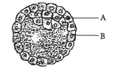
Question. (a) Name the human embryonic stage shown below. Identify ‘A’ and ‘B’ in it.
(b) Mention the part of the above embryonic stage that forms the foetus.
Answer :
(a) Blastocyst is shown in the figure. ‘A’ is inner cell mass and ‘B’ is trophoblast.
(b) Inner cell mass form the foetus.
Question. Mention the number of cells in the following stages

Answer : (a) 1 (b) 16 (c) 64
Question. Write the location and functions of the following in human testis :
(i) Sertoli cells
(ii) Leydig cells
Answer : (i) Location : Lined inside the seminiferous tubules.
Function : Provide nutrition to the germ cells / sperms.
(ii) Location : Outside seminiferous tubules / Interstitial space.
Function : Synthesize or secrete male hormones / Testicular hormones / Androgens.
Question. Differentiate between vas deferens and vasa efferentia.
Answer :
| S. No. | Vas deferens | Vasa efferentia |
| (i) | Carries sperm from epididymis to urethra. | Carries sperm from testis to epididymis. |
| (ii) | One in number from each testis. | Many in number. |
Detailed Answer :
| S. No. | Vas deferens | Vasa efferentia |
| (i) | They are long and curved tubes. | They are short and straight tubes |
| (ii) | They develop from cauda epididymis. | They develop from rete testis. |
| (iii) | They are broader and muscular. | They are narrower and delicate. |
| (iv) | They conduct sperm through muscular activity. | Sperm conduction takes place by ciliary current. |
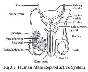
Question. Draw a labelled diagrammatic view of human male reproductive system.
Answer :
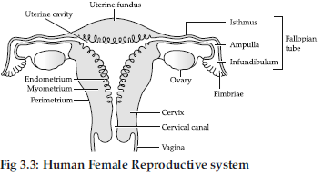
Question. Draw a labelled diagram of the human female reproductive system.
Answer :
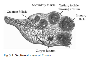
Question. Draw a sectional view of human ovary and label the different follicular stages, ovum and corpus luteum.
Answer :
Question. Draw a labelled diagrammatic sectional view of a human seminiferous tubule.
Answer :
Question. When do the oogenesis and spermatogenesis initiate in human females and males respectively ?
Answer : Oogenesis in females is initiated in the foetal or embryonic stage whereas spermatogenesis in males start at puberty. Sperm formation (spermatogenesis) in men continues even upto old age but formation of ovum in women ceases at about the age of 50 years with menopause.
Long Answer Type Questions
Question. (i) Draw a labelled diagrammatic view of human male reproductive system. R [Delhi, 2014]
(ii) Differentiate between :
(a) Spermatogenesis and spermiogenesis.
Answer :
| Spermatogenesis | Spermiogenesis |
| (i) It is the process of formation of aploid spermatozoa from germinal cells. | (i) It is the process of differentiation of spermatozoa from a spermatid. |
| (ii) A spermatogonium forms four spermatozoa. | (ii) Here, a spermatid forms a single spermatozoon. |
Question. The following is the illustration of the sequence of ovarian events “A” to “I” in a human female
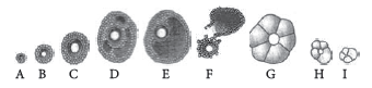
(a) Identify the figure that illustrates corpus luteum and name the pituitary hormone that influences its formation.
(b) Specify the endocrine function of corpus luteum. How does it influence the uterus? Why is it essential?
(c) What is the difference between “D” and “E”?
(d) Draw a neat labelled sketch of Graafian follicle.
Answer : (a) Figure ‘G’ illustrates corpus luteum.Rapid secretion of LH (luteinising hormone) from pituitary gland induces rupture of Graafian follicle and formation of corpus luteum.
(b) The corpus luteum secretes large amounts of progesterone and also estrogen. These hormones are essential for maintenance of the endometrium of uterus. Endometrium is necessary for implantation of fertilised ovum and the other events of pregnancy.
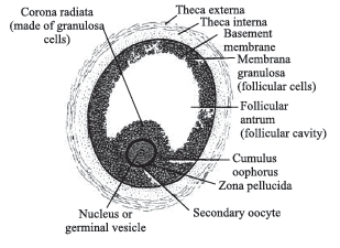
(c) Figure ‘D’ is tertiary follicle which is characterised by a fluid filled cavity called antrum.The theca layer is organised into an inner ‘theca interna’ and an outer ‘theca externa’. It is at this stage the primary oocyte within the tertiary follicle grows in size and completes its first meiotic division. It is an unequal division resulting in the formation of large haploid secondary oocyte and a tiny first polar body.Figure ‘E’ is mature follicle or Graafian follicle. In the Graafian follicle, the antrum enlarges further.
The secondary oocyte forms a new membrane called zona pellucida surrounding it.
(d) The diagram of Graafian follicle is as follows:
Question. Study the graph given below and answer the questions that follow :
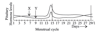
(a) Name the hormones ‘X’ and ‘Y’.
(b) Identify the ovarian phases during a menstrual cycle
(i) 5th day to 12th day of the cycle
(ii) 14th day of the cycle
(iii) 16th day to 25th day of the cycle
(c) Explain the ovarian events (i), (ii) and (iii) under the influence of hormones ‘X’ and ‘Y’.
Fertilisation and Implantation
Answer :
(a) X – Luteinising hormone (LH)
Y – Follicle stimulating hormone (FSH)
(b) (i) Proliferative phase
(ii) Ovulatory phase
(iii) Luteal phase
(c) (i) During follicular phase, FSH stimulates the ovarian follicle to secrete estrogen. Estrogen stimulates the proliferation of the endometrium of the uterine wall.
(ii) During ovulatory phase, both LH and FSH attain a peak level. Rapid secretion of LH induces rupturing of Graafian follicle and thereby the release of ovum.
(iii) During luteal phase, LH causes the remaining cells of the ovarian follicles to develop corpus luteum.
Question. When and where are primary oocytes formed in human female? Trace the development of these oocytes till ovulation (in menstrual cycle). How do gonadotropins influence this
developmental process?
Answer : Primary oocytes are formed inside the ovary during the embryonic developmental stage. Oogenesis is initiated during the embryonic development stage when oogonia are formed within each fetal ovary. These cells start division and enter into prophase-I of the meiotic division and get temporarily arrested at that stage, called primary oocytes. Each primary oocyte then gets surrounded by a layer of granulosa cells and is called the primary follicle. The primary follicles get surrounded by more layers of granulosa cells and a new theca and are called secondary follicles. The secondary follicle soon transforms into a tertiary follicle which is characterised by fluid filled cavity called antrum. The theca layer is organised into an inner theca interna and an outer theca externa. The primary oocyte within the tertiary follicle grows in size and completes its first meiotic division. It is an unequal division resulting in the formation of a large haploid secondary oocyte and a tiny first polar body. The tertiary follicle further changes into the mature follicle or Graafian follicle. The secondary oocyte forms a new membrane called zona pellucida surrounding it. The Graafian follicle now ruptures to release the secondary oocyte (ovum) from the ovary by the process called ovulation.
Effect of gonadotropins : Gonadotropin releasing hormone (GnRH) is secreted by the hypothalamus, which stimulates the release of follicle stimulating hormone (FSH) and luteinising hormone (LH).
FSH stimulates the ovarian follicles to produce estrogens, which stimulate the proliferation of the endometrium of uterine wall. LH causes ovulation and stimulates the corpus luteum of the ovary to secrete progesterone.
Question. Where does fertilisation occur in humans?
Explain the events that occur during this process.
Answer : The motile sperms swim rapidly, pass through the cervix, enter into the uterus and finally reach the junction of the isthmus and ampulla (ampullaryisthmic junction) of the Fallopian tube. The ovum released by the ovary is also transported to the ampullary-isthmic junction where fertilisation takes place. During fertilisation, a sperm comes in contact with the zona pellucida layer of the ovum and induces changes in the membrane that block the entry of additional sperms. The secretions of the acrosome help the sperm enter into the cytoplasm of the ovum through the zona pellucida and the plasma membrane.
This induces the completion of the meiotic division of the secondary oocyte. The second meiotic division is also unequal and results in the formation of a second polar body and haploid ovum (ootid). Soon the haploid nucleus of the sperms and that of the ovum fuse together to form a diploid zygote.
Question. List the different parts of the human oviduct through which the ovum travels till it meets the sperm for fertilisation.
Answer : Fimbriae, infundibulum, ampulla and isthmus are the main parts of oviduct, through which ovum travels till it meets the sperm for fertilisation. Finally it reach the ampullary-isthmic junction of oviduct.
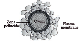
Question. Draw a diagram of the structure of a human ovum surrounded by corona radiata. Label the following parts :
(i) Ovum
(ii) Plasma membrane
(iii) Zona pellucida
Answer : The diagram of human ovum surrounded by corona radiata is as follows:
Question. Name the stage of the human embryo that gets implanted in the uterus and draw its labelled diagram.
Answer : Blastocyst gets implanted in the uterus

Question. Briefly explain the events of fertilisation and implantation in an adult human female.
Answer : The events of fertilisation in human female are:
(i) Acrosomal reaction : After ovulation, the secondary oocyte reaches the Fallopian tube. The capacitated sperm releases hydrolytic enzymes (sperm lysins) present in the acrosome, when it comes in contact with surface of egg covering.Important sperm lysins are (i) hyaluronidase that acts on the ground substances of follicle cells, (ii) corona penetrating enzyme that dissolves corona radiata and (iii) zona lysine or acrosin that helps to digest the zona pellucida. Due to acrosomal reaction, plasma membrane of sperm fuses with that of secondary oocyte and depolarisation of oocyte membrane occurs.
(ii) Cortical reaction : Immediately after the fusion of sperm and egg plasma membranes, the egg shows a cortical reaction to further check the entry of more sperms. In this reaction, the cortical granules present beneath the ovum’s plasma membrane fuse with the same and release their contents (enzymes) between it and zona pellucida. These enzymes harden the zona pellucida, which now functions as the sure block to polyspermy.
(iii) Sperm entry : The egg extends around the entering sperm, finger-like processes, called microvilli, which constitute a fertilisation cone. The latter take the entire sperm into the egg. The distal centriole of the sperm divides and forms two centrioles to generate the mitotic spindle for cell division.
(iv) Karyogamy : The sperm entry stimulates the egg (secondary oocyte) to resume and complete the suspended meiosis – II. This produces a haploid mature ovum and a second polar body. The head of sperm separates from the middle piece and tail to become male pronucleus and nucleus of ovum is called female pronucleus. The second polar body and sperm tail degenerate. Mixing up of the chromosomes of a spermatozoon and an ovum is called karyogamy or amphimixis. This completes the act of fertilisation. The fertilised ovum is now a diploid cell having 23 pairs of chromosomes, and is termed zygote.
The events of implantation are discussed as follows:
Implantation is the attachment of blastocyst to the uterine wall. It occurs after 7 days of fertilisation. As zygote moves towards the uterus, it undergoes series of mitotic divisions known as cleavage and forms 2,4,8,16 daughter cells called blastomeres. The embryo with 8 blastomeres is called morula. The morula transforms into blastocyst. In a blastocyst, the blastomeres are arranged into an outer layer called trophoblast and an inner group of cells called the inner cell mass. The trophoblast then gets attached to the endometrium and the inner cell mass gets differentiated as the embryo. After attachment the uterine cells divide rapidly and cover the blastocyst. As a result, the blastocyst becomes embedded in the endometrium of the uterus. This whole phenomenon is called implantation and it leads to pregnancy.
Question.
(a) One of the sperms is observed to penetrate ‘A’ of the ovum, as shown in the above diagram.Name ‘A’.
(b) How is the sperm able to do so?
(c) Where exactly in the Fallopian tube does this occur?
(d) Explain the events thereafter upto morula stage.
Answer : (a) A is zona pellucida.
(b) Sperm is able to penetrate the egg with the help of sperm lysins.
(c) Ampullary–isthmic junction.
(d) As soon as the sperm enters the egg, meiotic division of the secondary oocyte gets completed.
This results in the formation of a second polar body and a haploid ovum. Then, the haploid nucleus of the sperm and ovum fuse together to form a diploid zygote. The mitotic division starts as the zygote moves through the oviduct called cleavage towards the uterus and forms daughter cells called blastomeres. The embryo with 8 to 16 blastomeres is called a morula.
Question. Where is morula formed in humans? Explain the process of its development from zygote.
Answer :
Morula is formed inside the oviduct. As the zygote moves through isthmus of the oviduct towards the uterus, mitotic divisions called cleavage starts. The daughter cells so formed are called blastomeres. The embryo with 8 to 16 blastomeres is called morula.
Pregnancy and Embryonic Development
Question. Comment on the role of placenta as an endocrine gland.
Answer : The placenta acts as an endocrine gland and secretes the following hormones :
(i) Human chorionic gonadotropin (hCG)
(ii) Human chorionic somatomammotropin (hCS)
(iii) Progesterone (iv) Estrogen (v) Relaxin
(vi) Chorionic thyrotropin and (vii) Chorionic corticotropin
The hCG stimulates and maintains the corpus luteum to secrete progesterone until the end of pregnancy. The hCS stimulates the growth of the mammary glands during pregnancy. Relaxin facilitates parturition (act of birth) by softening the connective tissues of the pubic symphysis. The level of hormones like estrogen, progesterone etc. are increased in maternal blood during pregnancy. Increased production of these hormones is necessary for supporting the fetal growth, metabolic changes in mother and maintenance of pregnancy.
Question. When and where do chorionic villi appear in humans? State their function.
Answer : After implantation, finger-like projections appear on the trophoblast called chorionic villi which are surrounded by the uterine tissue and maternal blood. The chorionic villi and uterine tissue become interdigitated with each other and jointly form a structural and functional unit between developing embryo (foetus) and maternal body called placenta, which facilitates the supply of oxygen and nutrients to the embryo and also removal of carbon dioxide and excretory waste materials produced by the embryo. Placenta also acts as an endocrine tissue and produces several hormones essential for supporting the foetal growth, metabolic changes in the mother and maintenance of pregnancy.
Question. Explain the function of umbilical cord.
Answer : The placenta is connected to embryo through an umbilical cord which helps in the transport of substances to and from the embryo.
Question. Explain the fate of inner cell mass in a human embryo immediately after implantation.
Answer : Immediately after implantation, the inner cell mass (embryo) differentiates into an outer layer called ectoderm and an inner layer called endoderm. A mesoderm soon appears between the ectoderm and the endoderm. These three layers give rise to all tissues (organs) in adults.
Question. Write the function of oxytocin.
Answer : Oxytocin acts on the uterine muscle causing their contractions that help in the expulsion of the baby out of the uterus through the birth canal.
Question. What stimulates pituitary to release the hormone responsible for parturition? Name the hormone.
Answer : THe signals from the fully developed foetus and placenta triggers release of oxytocin from the maternal pituitary. Oxytocin is responsible for parturition.
Question. Why is oxytocin called as ‘birth hormone’ ?
Answer : Oxytocin is called as ‘birth hormone’ as it causes increased contraction of the uterine muscles and thus explusion of the baby from the uterus through the birth canal.
Question. (a) Where do the signals for parturition originate from in humans?
(b) Why is it important to feed the newborn babies on colostrum?
Answer :(a) The signals for parturition originate from the fully developed foetus and the placenta which induce mild uterine contractions called foetal ejection reflex.
(b) After birth, the breast first release milk is called colostrum for 2 or 3 days. This is a thin, yellowish, fluid, often called foremilk. It contains cells from the alveoli and is rich in protein (lactalbumin & lactoprotein), antibodies, but low in fat. Breast-feeding during the initial period of infant growth is recommended by doctors for bringing up a healthy baby.
Question. Why is parturition called a neuroendocrine mechanism?
Answer : Process of parturition is induced by both neural system and endocrine system therefore, it is called a neuro-endocrine mechanism. The signals for parturition originate from the fully developed foetus and the placenta which induce mild uterine contractions called foetal ejection reflex. This triggers release of oxytocin from the maternal pituitary. Oxytocin acts on the uterine muscle and causes stronger uterine contractions that leads to expulsion of the baby out of the uterus through the birth canal.
Question. Why is breast feeding recommended during the initial period of an infant’s growth? Give reason.
Answer : Secretion and storage of milk generally begins after birth of young one, usually within 24 hours under the influence of hormone prolactin (PRL) secreted by anterior lobe of the pituitary gland. However, the ejection of milk is stimulated by the hormone oxytocin (OT) released from the posterior lobe of the pituitary gland.
Question. Describe the process of parturition in humans.
Answer : The act of expelling the full term young one from the mother’s uterus at the end of gestation period is called parturition. Process of parturition is induced by both nervous system and hormones secreted by the endocrine glands of the mother. The signals for child birth (parturition) originate from the fully matured foetus and placenta which induce mild uterine contractions called foetal ejection reflex. This causes quick release of oxytocin from the maternal pituitary gland.
Oxytocin acts on the uterine muscle and causes stronger uterine contractions which in turn further stimulates the secretion of oxytocin. The stimulatory reflex between the uterine contraction and oxytocin secretion continues resulting in stronger and stronger contractions. This leads to expulsion of the baby from the uterus through the birth canal.